Science keeps evolving. In life sciences, researchers no longer just run tests and write notes. They want to actually see what happens at the tiniest levels. That’s where imaging steps in. It opens up new ways to explore cells, molecules, and the processes that drive them. These days, images do more than support data—they are data.
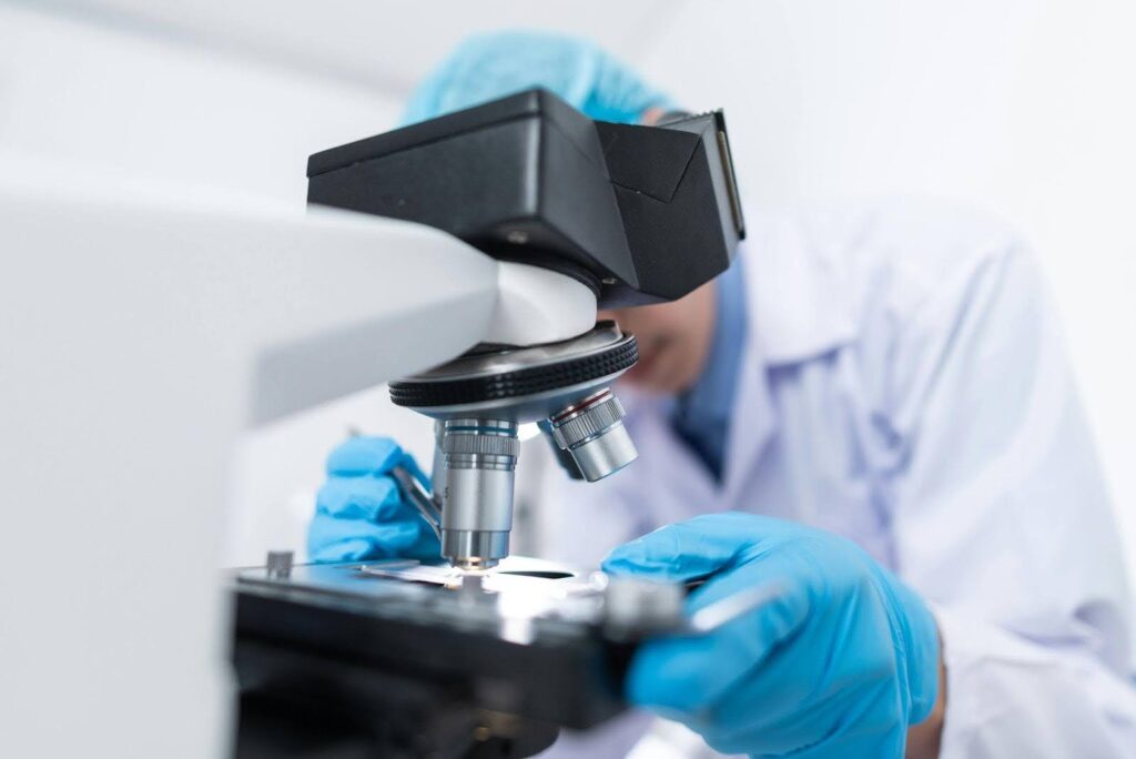
One popular tool in many research labs is the fluorescence microscope. It allows scientists to highlight specific parts of a cell using special dyes. These dyes glow when exposed to certain light. That glow helps researchers zero in on details they couldn’t spot before. It becomes easier to follow where a protein moves or how a cell reacts under stress.
Looking at the Fine Points
Precision imaging means more than just getting clear pictures. It’s about collecting useful details from those pictures. If a researcher can see what’s happening in real time, they’re more likely to find answers. Regular microscopes don’t always show enough. Precision tools can spot tiny shifts in shape, size, or motion.
This makes a huge difference in research. A scientist can watch how a virus spreads. They can measure how fast a cell divides. They can even see what changes first when a drug is applied. These images don’t just look good—they give direction. They guide the next step.
Letting Images Speak
Today, a single image can carry a lot of meaning. But a full set of images tells a story. Precision imaging allows researchers to track what’s going on over time. This builds a deeper picture of how systems behave. With the help of smart software, those visuals turn into numbers, charts, and insights.
For example, a scientist may observe how a group of cells responds to a new chemical. They can count how many cells survive. They can see changes in shape or behavior. And they can compare the results across several tests. All of this is pulled from image data.
Watching Cells in Real Time
Live-cell imaging is another game-changer. Instead of working with dead samples, researchers can now watch living cells as things happen. They no longer need to stop the process just to take a closer look. They can stay with it.
That’s very useful during experiments with new treatments. Scientists can add a substance and instantly see how cells react. They can spot early warning signs. They can record recovery or failure. This helps speed up testing and cuts down on wasted effort.
Smarter Tools for Faster Work
Imaging today isn’t just about the camera. It’s also about what happens after the photo is taken. Artificial intelligence now plays a role in scanning, sorting, and analyzing images. This speeds up the process and helps reduce missed details.
AI can look through thousands of files in a short time. It highlights patterns. It flags problems. It even suggests next steps. That lets researchers focus on what matters. They can work with the key images and ignore the noise.
Not Just for Human Cells
Imaging helps outside the lab, too. In farming, for instance, researchers use it to study how roots grow. They track how plants handle heat, pests, or poor soil. This helps improve crops and plan for climate changes.
In environmental science, imaging allows people to study tiny creatures like algae or microbes. These organisms help clean up waste or break down harmful materials. By watching their behavior, scientists learn how to improve cleanup systems. Imaging supports that kind of problem-solving.
Some Limits Still Remain
As useful as imaging is, it comes with challenges. Equipment can be pricey. Some labs just don’t have the funds for it. Even when they do, they need trained people to run the machines. That takes time and effort.
Another issue is data. High-quality images take up a lot of space. They require powerful computers and special software to process. Not every lab is set up for that. Still, many groups find ways around these problems. They team up with other labs. They use cloud storage. They turn to open-source tools. With the right plan, even small teams can make it work.

What’s Next?
Precision imaging keeps moving forward. New technologies are on the way. Some offer faster scans. Others build 3D models of cells or tissues. Some even combine multiple imaging types into one system. These upgrades will bring better tools to more researchers.
This progress means more than better pictures. It means deeper understanding. It helps scientists find new answers. It supports faster breakthroughs. And it gives the life sciences more ways to explore problems and test solutions. The better we see, the more we learn.